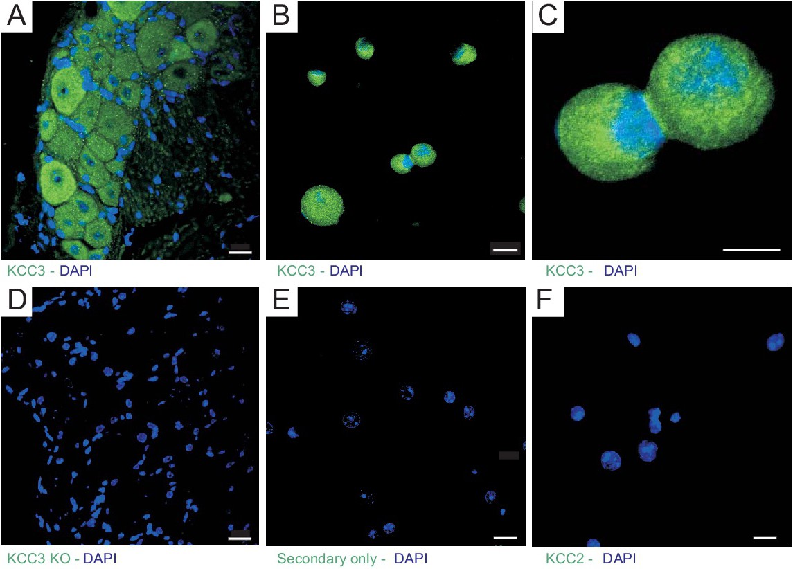
Fig. 1. Expression of KCC3 in mouse DRG neurons. A, Section of dorsal root ganglion isolated from wild-type mice stained with rabbit anti-KCC3 polyclonal antibody, followed by FITC-conjugated anti-rabbit antibody. B-C, DRG neurons isolated from wild-type mice stained with rabbit anti-KCC3 polyclonal antibody followed by FITC-conjugated anti-rabbit antibody. D, Section of dorsal root ganglion isolated from KCC3 knockout mice, stained with rabbit anti-KCC3 polyclonal antibody, followed by FITC-conjugated anti-rabbit antibody. E, Isolated dorsal root ganglion neurons stained with secondary antibody only. F, wild-type DRG neurons stained with rabbit anti-KCC2 polyclonal antibody show absence of KCC2 expression. All slides were mounted with DAPI-containing mounting reagent to stain nuclei. Bars: 20 µm for A, B, D-F and 10 µm for C.